
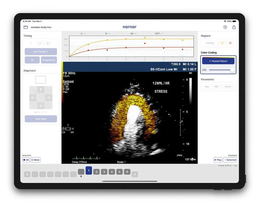
The free narnar app for CEUS allows researchers to process, visualize and quantify perfusion studies of various organs (e.g., heart, kidney, liver, skeletal muscle). The app supports a range of research protocols to meet diverse research needs.
Try the narnar app for research in contrast-enhanced ultrasound.
Key Features
Automated and Manual Alignment
Averaging
MIP Imaging

Digital Subtraction and Color Coding

Parametric Perfusion Imaging

Open DICOM Files
Trusted by Hundreds of Researchers Worldwide
Our research software has been widely used for contrast-enhanced ultrasound research. Selected institutions include:
Oregon Health & Science University
University of Virginia
Mayo Clinic
Brigham and Women's Hospital
National Institutes of Health
Zhejiang University
For the full list, click here.
FAQs
-
The narnar app is designed for iPad Pro (11 inch, 12.9-inch, and 10.5-inch models are supported). We highly recommend the Apple Pencil and Smart Keyboard. The app will still work without them, but you will not be able to fully enjoy the full usability those tools provide.
How it works:get an iPad Pro + keyboard + pencil,
download the free version of narnar app from the App Store,
start using the app, let us know at support@narnar.co if you need more help,
let us know if you are interested in participating in our beta program.
-
DICOM format is limitedly supported for reading files generated by systems from major ultrasound vendors.
-
Once you have the app installed, simply take a screenshot by pressing the home and power buttons. You will be prompted to submit feedback by email. Or contact us with your suggestions or bug reports directly at support@narnarhealth.com
-
Ask us about our beta program if you are interested in helping us to develop new features.
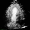
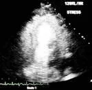
Examples (Cardiology)

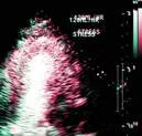
Digital subtraction and color coding developed using our software contributed to the advancements in CEUS since its early years.

Perfusion Defect: High MI Imaging of AMI, alignment, averaging.

Averaging to improve myocardium delineation

Normal Perfusion: Low MI Imaging and quantification of the heart
Activated Droplets: Infarct zone imaging
Examples (Other Organs)
![[Kidney] Normal kidney perfusion, images aligned and averaged.](https://images.squarespace-cdn.com/content/v1/68926c61b505126b7e78f27f/21df7b9a-43f5-462c-b402-2bf5c8868b17/screenshot-2019-03-02-23-16-20.png)
[Kidney] Normal kidney perfusion, images aligned and averaged.
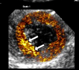
![[Kidney]](https://images.squarespace-cdn.com/content/v1/68926c61b505126b7e78f27f/f112e8dd-f483-4bb9-92ac-8c6ee2a92585/kidney-for-web.png)
[Kidney]
![[Liver] Focal nodular hyperplasia (FNH), a benign tumor-like mass of the liver.](https://images.squarespace-cdn.com/content/v1/68926c61b505126b7e78f27f/cc8cf4fb-bb04-4ed7-b52b-334f02e99e20/liver-sequence.jpg)
[Liver] Focal nodular hyperplasia (FNH), a benign tumor-like mass of the liver.
![[Placenta] Gradient of blood flow in a placental cotyledon.](https://images.squarespace-cdn.com/content/v1/68926c61b505126b7e78f27f/202a8ff0-75f7-4af6-81ce-897b61329389/placenta.jpg)
[Placenta] Gradient of blood flow in a placental cotyledon.
![[Uterus] Averaged](https://images.squarespace-cdn.com/content/v1/68926c61b505126b7e78f27f/820ef7c0-c90c-47db-b701-7bd3053a6adf/uterusaveraged-otherfocus-web.jpg)
[Uterus] Averaged
![[Uterus] Parametric image of blood flow velocity.](https://images.squarespace-cdn.com/content/v1/68926c61b505126b7e78f27f/130ecc1e-c4b2-44c0-b8f7-2acbdb63117c/uterusbetaparam-web.jpg)
[Uterus] Parametric image of blood flow velocity.
![[Skeletal Muscle] MIP images are enhanced by color-coding.](https://images.squarespace-cdn.com/content/v1/68926c61b505126b7e78f27f/58a78086-70e2-45d2-bdb0-7945fd0f97eb/splitmuscle.png)
[Skeletal Muscle] MIP images are enhanced by color-coding.
![[Brain] Parametric image of blood flow in a small animal model.](https://images.squarespace-cdn.com/content/v1/68926c61b505126b7e78f27f/f774a78f-2efe-4ad0-ac55-66ad9c8ef378/brain.png)
[Brain] Parametric image of blood flow in a small animal model.
Contact us.
support@narnarhealth.com